- Home/
- Technology/
- Structural Variation


Structural Variation Detection
Bionano’s genome mapping technology directly observes long molecules from 150,000 bp to megabase pair lengths to identify large structural variations typically not detected by short- or long-read sequencing technologies.
Sequence Variation Identification
Identification of sequence variation is an important first step in determining critical genetic components of a phenotype. Structural variations (SVs) impact large stretches of sequence and are likely to impact phenotype.
Retaining long-range contiguity throughout the genome mapping process is critical for any comprehensive study of genome structure and function, particularly for the analysis of structural variation in complex genomes. Bionano genome mapping offers unmatched sensitivity for the detection of large SVs.1
- 99% sensitivity for homozygous insertions/deletions larger than 500 base pairs
- 95% sensitivity for heterozygous insertions/deletions larger than 500 base pairs
- 95% sensitivity for balanced and unbalanced translocations larger than 50,000 base pairs
- 99% sensitivity for inversions larger than 30,000 base pairs
- 97% sensitivity for duplications larger than 30,000 base pairs
- 97% sensitivity for copy number variants larger than 500,000 base pairs
For mosaic samples or heterogeneous cancer samples, Saphyr detects all types of structural variants down to 1% Variant Allele Fraction.
To identify a structural variation, a de novo genome map assembly can be aligned to a reference genome, or two samples can be aligned to each other directly. When aligning a genome map to a reference assembly, Bionano software identifies the location of the same recognition sequence used to label the DNA molecules in the reference genome and aligns matching label patterns in the sample and reference. This alignment provides all the annotation of the reference to the de novo assembled genome.
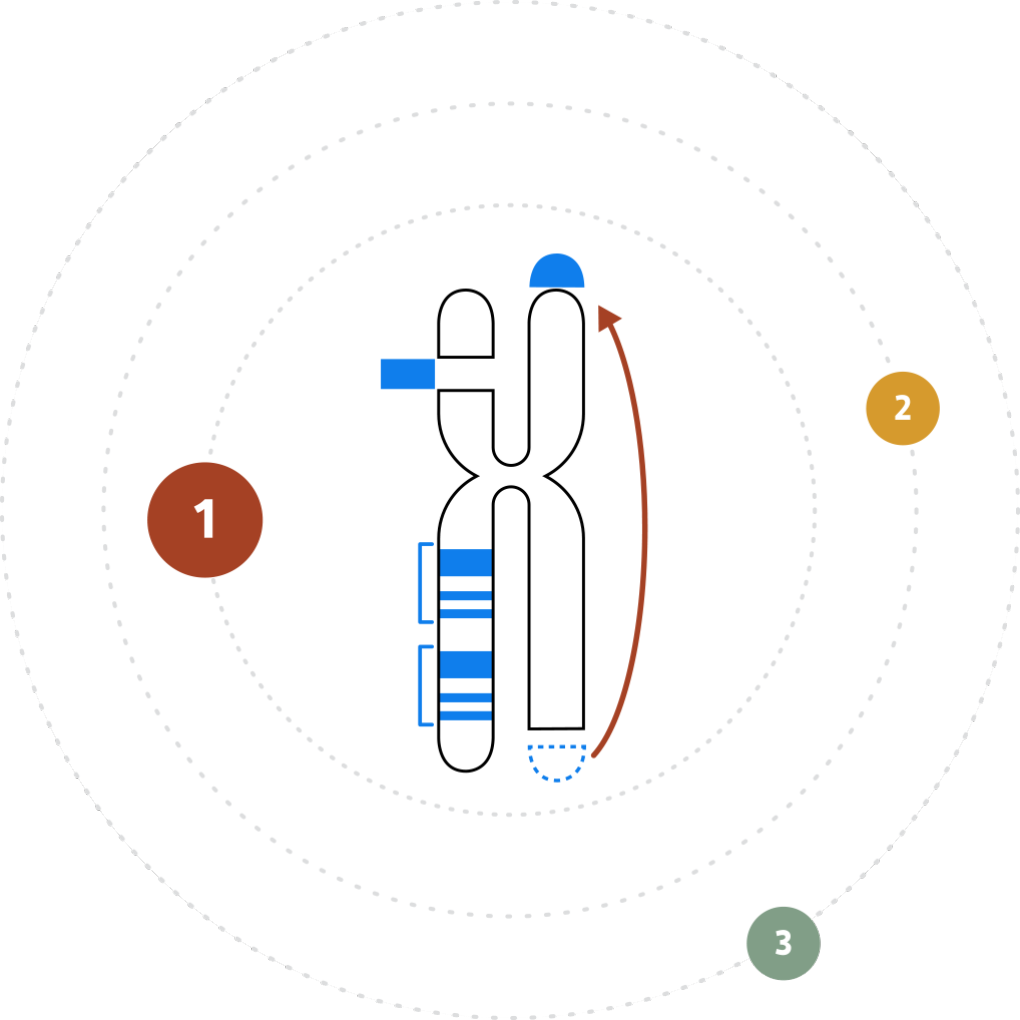
By observing changes in label spacing and comparisons of order, position, and orientation of label patterns, Bionano’s automated structural variation calling algorithms detect all major structural variation types.

A decreased spacing of labels, with or without loss of labels, is evidence of deletions. Label spacing that increases with or without additional labels detected point to inserted sequences.
Using similar methods, expansions or contractions of tandem arrays or segmental duplications can be identified. When label patterns are inverted relative to the reference, an inversion is called. Genome maps aligning partially with two or more different chromosomes or genomic locations indicate translocations.
Family-Based and Case-Control Studies
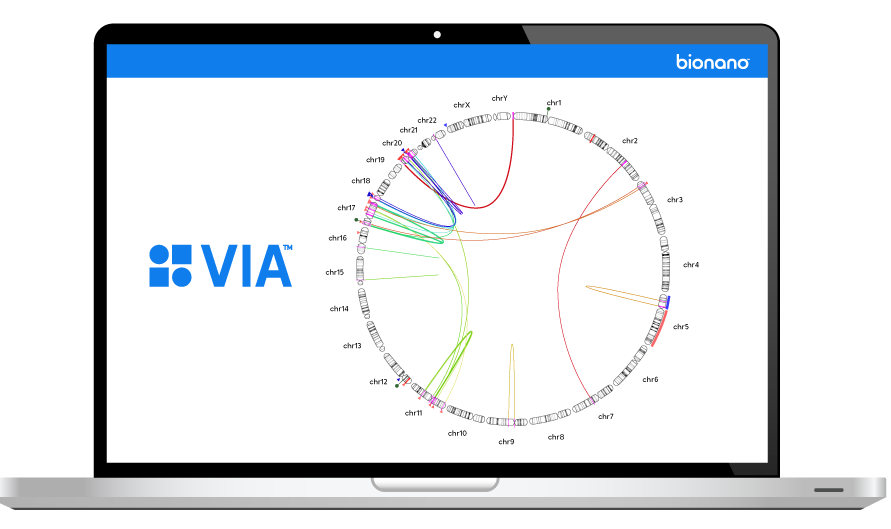
Bionano’s Variant Annotation Pipeline (VAP) streamlines family-based and case-control studies. Using VAP, structural variation calls from multiple samples can be analyzed together to detect inherited and de novo structural variations. The pipeline does so by comparing SV calls from the parent(s) with the child, or by comparing structural variation calls from the tumor with the blood from the same patient to detect somatic mutations.
By using a control database of common structural variations, VAP filters the thousands of identified down to hundreds of rare variants, or a handful of de novo variants and the genes they affect. The structural variation calling and VAP are fully integrated with Bionano Access™, which provides a convenient interface for running and viewing structural variation analysis results.
Structural Variation Analysis with Bionano
With Bionano, structural variations are observed, not inferred as they are with NGS. When short-read NGS sequences are aligned to the reference genome, algorithms piece together sequence fragments in an attempt to rebuild the actual structure of the genome. In this approach, structural variations are inferred from the fragmented data with mixed success. With Bionano, megabase-size native DNA molecules are imaged, and most large structural variations or their breakpoints can be observed directly in the label pattern on the molecules.
Learn more about Bionano Data Solutions.