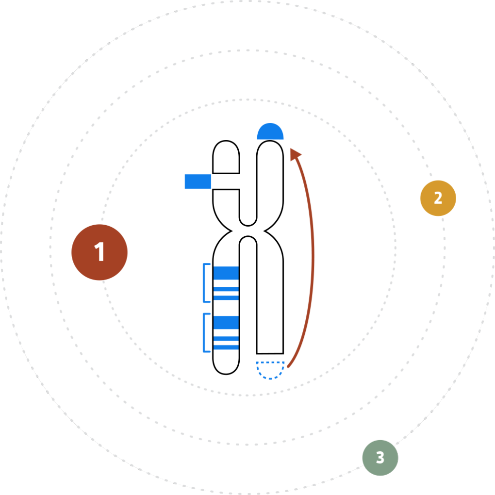
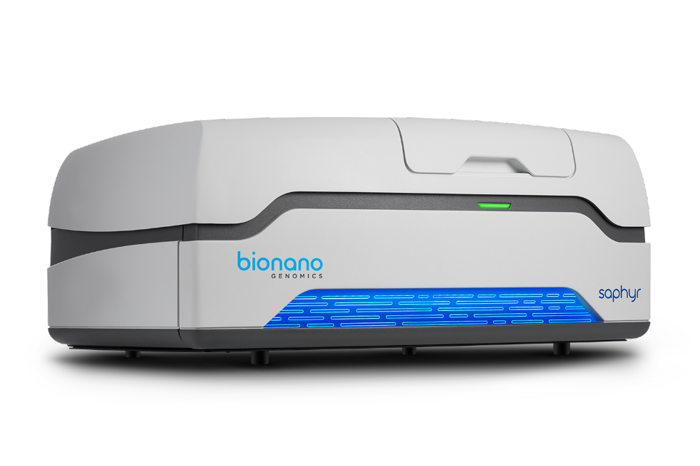
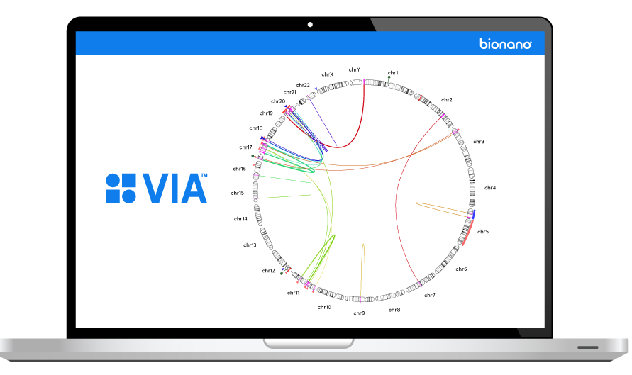
The Bionano Genome Imaging workflow starts with mega-base size DNA isolation. A single enzymatic reaction labels the genome at a specific sequence motif occurring approximately 15 times per 100 kbp in the human genome. The long, labeled DNA molecules are linearized in nanochannel arrays on a Saphyr chip® and imaged in an extremely high throughput, automated manner by the Saphyr® Genome Imaging Instrument. Using pairwise alignments, the molecules are assembled into local maps or whole genome de novo assemblies. Changes in patterning or spacing of the labels are detected automatically, genome wide, to call all structural variants.
- Home/
- Discovery Research /
- Genetic Diseases
Genetic Disease
Research
Detect structural variations, unbiased and genome-wide with Bionano optical genome mapping at high sensitivities and extremely low false positive rates. Discover the variants that are missed by short and long read sequencing with one powerful workflow.
Bionano finds the variants NGS can’t see
Structural variants (SVs) make up the majority of human genomic variation, driving genetic diversity but contributing to genetic diseases. SVs are frequently flanked by repetitive sequences which are challenging or impossible for current sequencing technologies to decipher. Bionano optical genome mapping reveals SVs with up to 99% sensitivity, even at allele fractions as low as 1%, which is not possible using other genomics technology.
Data Examples
Many protein-coding exons are ‘camouflaged’ in NGS datasets because of variably-repeated binding domains—the exons occur in more than one gene or in tandem within the same gene, making correct alignment of short reads impossible. Saphyr allows for the direct measurement of the number of C3b/C4b binding domains for each haplotype in CR1, an Alzheimer associated gene, in this patient with Alzheimer’s Disease.
In a newborn with Congenital Diaphragmatic Hernia (CDH), a severe developmental disorder affecting the diaphragm, lungs and sometimes heart, Bionano detected two adjacent duplications, one direct and one inverted. Bionano revealed a much more complex architecture than could be inferred from microarray data and identified several additional candidate genes for CDH.
In a single postmortem brain sample from an ALS patient, Bionano Saphyr detected a highly mosaic range of expansions of the C9orf72 GGGGCC repeat, ranging from the reference allele to a 32 kbp expansion. No modern technology has been capable of spanning and measuring these large C9orf72 repeat expansions, but Bionano can.
In a patient with Duchenne Muscular Dystrophy (DMD), a 420 kbp segment from chromosome 15 was duplicated in an inverted orientation in intron 44 of the Dystrophin gene. This insertion was not detected by NGS, and while chromosomal microarray can detect the duplication, its location and therefore implication in DMD could not be determined.
Data Examples
The Bionano Genome Imaging workflow starts with mega-base size DNA isolation. A single enzymatic reaction labels the genome at a specific sequence motif occurring approximately 15 times per 100 kbp in the human genome. The long, labeled DNA molecules are linearized in nanochannel arrays on a Saphyr chip® and imaged in an extremely high throughput, automated manner by the Saphyr® Genome Imaging Instrument. Using pairwise alignments, the molecules are assembled into local maps or whole genome de novo assemblies. Changes in patterning or spacing of the labels are detected automatically, genome wide, to call all structural variants.

Many protein-coding exons are ‘camouflaged’ in NGS datasets because of variably-repeated binding domains—the exons occur in more than one gene or in tandem within the same gene, making correct alignment of short reads impossible. Saphyr allows for the direct measurement of the number of C3b/C4b binding domains for each haplotype in CR1, an Alzheimer associated gene, in this patient with Alzheimer’s Disease.

In a newborn with Congenital Diaphragmatic Hernia (CDH), a severe developmental disorder affecting the diaphragm, lungs and sometimes heart, Bionano detected two adjacent duplications, one direct and one inverted. Bionano revealed a much more complex architecture than could be inferred from microarray data and identified several additional candidate genes for CDH.

In a single postmortem brain sample from an ALS patient, Bionano Saphyr detected a highly mosaic range of expansions of the C9orf72 GGGGCC repeat, ranging from the reference allele to a 32 kbp expansion. No modern technology has been capable of spanning and measuring these large C9orf72 repeat expansions, but Bionano can.

In a patient with Duchenne Muscular Dystrophy (DMD), a 420 kbp segment from chromosome 15 was duplicated in an inverted orientation in intron 44 of the Dystrophin gene. This insertion was not detected by NGS, and while chromosomal microarray can detect the duplication, its location and therefore implication in DMD could not be determined.

While deletions are somewhat easier to detect by NGS, insertions are rarely picked up from NGS data. In a genetic male patient with gonadal dysgenesis, Bionano identified a 6 kbp insertion in the WDR11 gene, associated with abnormal testes development and cryptorchidism.

Related Materials


Optical genome mapping using Saphyr® reveals what’s missing in your research. Rapidly identify genome variation like never before with the high-throughput Saphyr system.
Built using proprietary Nanochannel technology, Bionano Chips for the Saphyr® and Irys® systems linearize DNA, enabling high-speed, high-throughput optical genome mapping and structural variation detection for a variety of applications including human and clinical research.
Bionano Prep Kits™ provide the critical reagents and protocols needed to extract and label high molecular weight (HMW) DNA for use on the Irys® and Saphyr™ systems. Bionano kits are optimized for performing Bionano optical genome mapping applications on a variety of sample types.